PARTS OF AN EYE
● `color{violet}("Our paired eyes")` are located in sockets of the `color{violet}("skull")` called `color{brown}("orbits.")`
● The `color{violet}("adult human eye ball")` is nearly a `color{brown}("spherical")` structure.
● The `color{violet}("wall of the eye ball")` is composed of three layers.
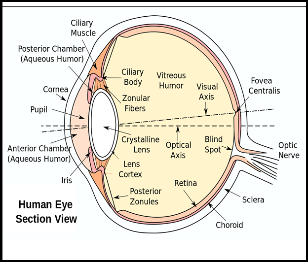
● The `color{violet}("external layer")` is composed of a `color{violet}("dense connective tissue")` and is called the `color{brown}("sclera. ")`
● The `color{violet}("anterior portion")` of this layer is called the `color{brown}("cornea. ")`
● The middle layer, `color{brown}("choroid,")` contains many `color{violet}("blood vessels")` and looks `color{violet}("bluish in colour. ")`
● The `color{violet}("choroid layer")` is thin over the posterior two-thirds of the `color{violet}("eye ball,")` but it becomes thick in the anterior part to form the `color{brown}("ciliary body.")`
● The `color{violet}("ciliary body")` itself continues forward to form a pigmented and opaque structure called `color{violet}("the iris")` which is the visible coloured portion of the `color{violet}("eye.")`
● The `color{violet}("eye ball")` contains a transparent `color{brown}("crystalline lens")` which is held in place by ligaments attached to the `color{violet}("ciliary body.")`
● In front of the `color{violet}("lens")`, the aperture surrounded by the iris is called the `color{brown}("pupil.")`
● The diameter of the `color{violet}("pupil")` is regulated by the `color{violet}("muscle fibres")` of iris.
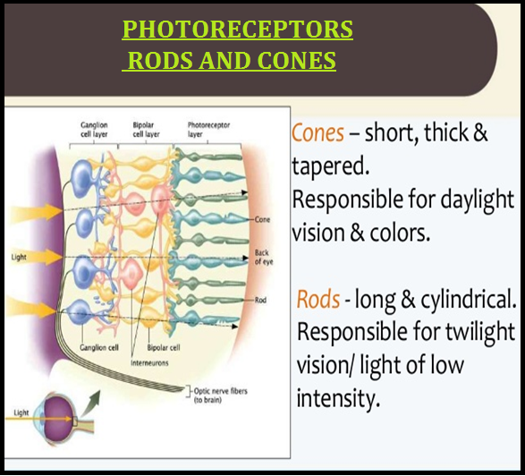
● The inner layer is the `color{brown}("retina")` and it contains three layers of cells – from inside to outside – `color{violet}("ganglion cells,")`
● The `color{violet}("adult human eye ball")` is nearly a `color{brown}("spherical")` structure.
● The `color{violet}("wall of the eye ball")` is composed of three layers.
● The `color{violet}("external layer")` is composed of a `color{violet}("dense connective tissue")` and is called the `color{brown}("sclera. ")`
● The `color{violet}("anterior portion")` of this layer is called the `color{brown}("cornea. ")`
● The middle layer, `color{brown}("choroid,")` contains many `color{violet}("blood vessels")` and looks `color{violet}("bluish in colour. ")`
● The `color{violet}("choroid layer")` is thin over the posterior two-thirds of the `color{violet}("eye ball,")` but it becomes thick in the anterior part to form the `color{brown}("ciliary body.")`
● The `color{violet}("ciliary body")` itself continues forward to form a pigmented and opaque structure called `color{violet}("the iris")` which is the visible coloured portion of the `color{violet}("eye.")`
● The `color{violet}("eye ball")` contains a transparent `color{brown}("crystalline lens")` which is held in place by ligaments attached to the `color{violet}("ciliary body.")`
● In front of the `color{violet}("lens")`, the aperture surrounded by the iris is called the `color{brown}("pupil.")`
● The diameter of the `color{violet}("pupil")` is regulated by the `color{violet}("muscle fibres")` of iris.
● The inner layer is the `color{brown}("retina")` and it contains three layers of cells – from inside to outside – `color{violet}("ganglion cells,")`